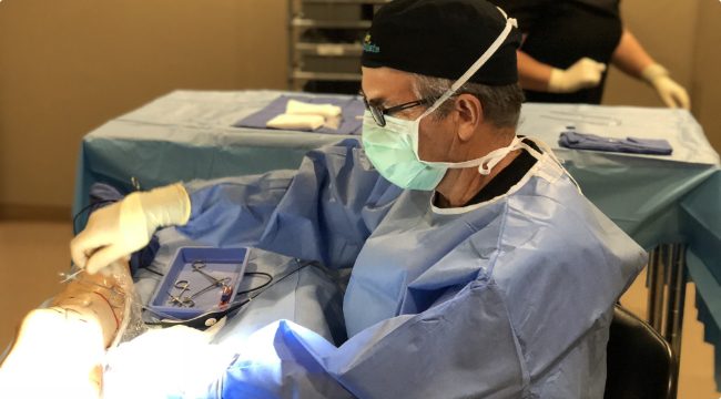
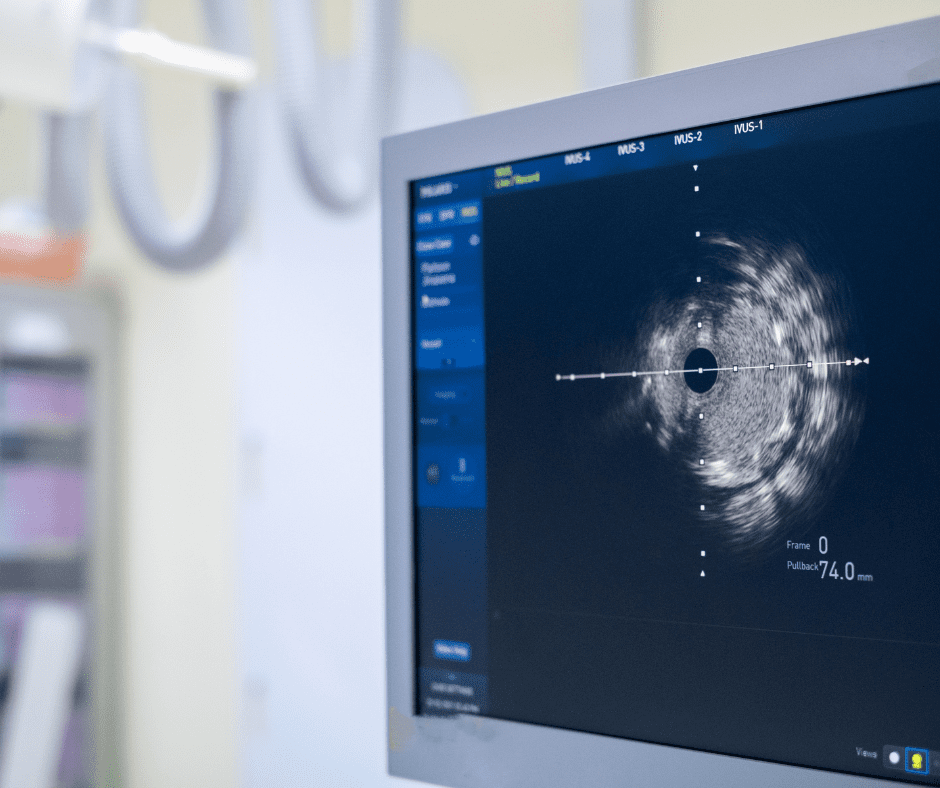
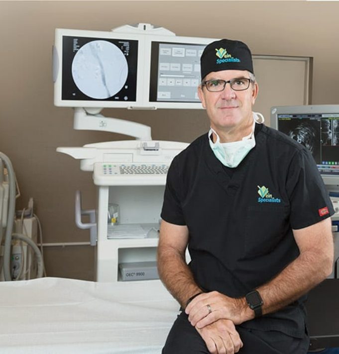
Intravascular ultrasound (IVUS) is a medical imaging methodology using a specially designed catheter with a miniaturized ultrasound probe attached to the patient end of the catheter with the other end attached to computerized ultrasound equipment. The end with the probe is inserted through an IV in the leg and directed into the pelvic veins of interest. IVUS allows for examination of the veins from within to check for obstructions. Precise localization and accurate measurement of the severity of the vein narrowing is possible with IVUS. Although stent technology and IVUS have been used for years in other vascular beds, the use of them together to investigate the pelvic veins is new and exciting in that many desperate vein patients as well as patients with undiagnosed pelvic and buttock pain previously evaluated by their OB/GYN or Pain Management physicians now have the potential of being helped.
Intravascular ultrasound as it applies to the venous system is focused on the identification of iliac vein compression syndrome (IVCS). One form of this is referred to as May-Thurner syndrome wherein the left common iliac vein is compressed between the crossing right common iliac artery and the 5th Lumbar vertebral body or the sacrum. The chronic, pulsatile compression of the proximal left common iliac vein by the right common iliac artery against the spine leads to peri-venous fibrosis or scarring. Blood flow from the legs has to find alternative routes of return through the pelvis to the heart, and these pelvic collateral veins result in the condition of pelvic venous congestion. Left sided lesions account for 75% of these cases while right sided iliac vein compression lesions account for the remaining 25% of cases seen all of which may benefit from IVUS evaluation and treatment. Thus, it is imperative to examine both iliac venous systems routinely when investigating for iliac vein compression. For example, a left sided iliac vein compression may present with right sided leg signs and symptoms and the left sided symptoms may (less frequently) be caused by right sided iliac vein compression lesions.
The resultant increased lower extremity venous hypertension due to the pelvic venous obstruction from iliac vein compression lesions may lead to significant thigh and leg swelling, failure of previous vein procedures with recurrent varicose veins, skin changes around the lower leg and ankle region and in some cases, non-healing venous ulcers. Patients may complain of left hip, flank or buttock pain, achiness down the posterior thigh into the posterior knee area. Women (and men more infrequently) may present with symptoms and signs of pelvic congestion syndrome, or may have been given the diagnosis of other obscure causes of their pelvic complaints, such as psychogenic pelvic pain or post-partum depression. IVUS allows precise identification and stratification of these blockages from within the iliac veins. This makes it possible to determine which patients are most likely to benefit significantly from IVUS directed therapy with balloon angioplasty and intravascular stent placement. Angioplasty (iliac vein venoplasty) stretches the scar tissue while the stent is required to maintain patency as the incidence of recoil and recurrent symptoms is too great without stent placement in this group of patients.