
Very superficial extrafascial parallel axial vein – foam closure vs surgical removal
Clinical Context
A young, very healthy, athletic woman presented after prior left-leg venous
treatment performed elsewhere.
Duplex demonstrated residual reflux feeding a very superficial, long, linear tributary
that tracked parallel to the expected course of the great saphenous vein (GSV).
The target vein appeared extrafascial (outside the saphenous fascia), consistent with an
accessory/parallel axial tributary rather than a true intrafascial GSV segment (Figure 1).
Initial decision and treatment.
Because the vein was immediately subdermal, I felt thermal ablation (EVLT/RFA) carried
an unacceptably high risk of skin injury. I therefore chose Varithena (polidocanol endovenous
microfoam) under ultrasound guidance.
Outcome and Problem
The treatment successfully closed the target vein; however, closure resulted in a sclerosed/thrombosed,
hyperpigmented, tender linear cord that was cosmetically conspicuous and painful (Figure 1).
Despite counseling and a prolonged period of observation, the patient remained dissatisfied.
Revision Procedure
After months of conservative management and shared decision-making, I returned and performed
surgical excision/microphlebectomy of the thrombosed superficial vein.
The re-exploration was technically challenging due to dense scarring and adherence, requiring
multiple small incisions; the thrombosed segment was removed in continuity (Figure 2 and
Figure 3).
Call Or Request An Appointment
Contact us to schedule an appointment with our expert Vein Specialists team. We will evaluate your signs and symptoms, answer your questions, and create a personalized vein care treatment plan to relieve your leg pain and enhance your life.
Schedule Your Appointment TodayTeaching Point
In patients with varicosities that are extremely superficial (near-dermal) – particularly long, linear,
parallel axial veins in the extrafascial space – surgical removal (microphlebectomy/excision) is
often preferable to foam sclerotherapy (including Varithena) or other ultrasound-guided
sclerotherapy.
Even when chemical closure is technically successful, it may leave a painful,
visible thrombosed/sclerosed cord with prolonged tenderness and hyperpigmentation.
Note: Images are clinician-supplied and intended for educational discussion.
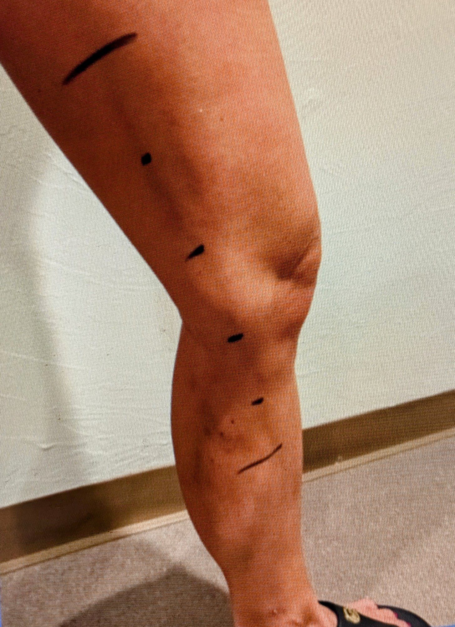
Figure 1. Pre-/interval appearance with marked superficial parallel axial vein course;
post-foam closure resulted in a visible, tender, near-dermal thrombosed/sclerosed cord
along the marked path.
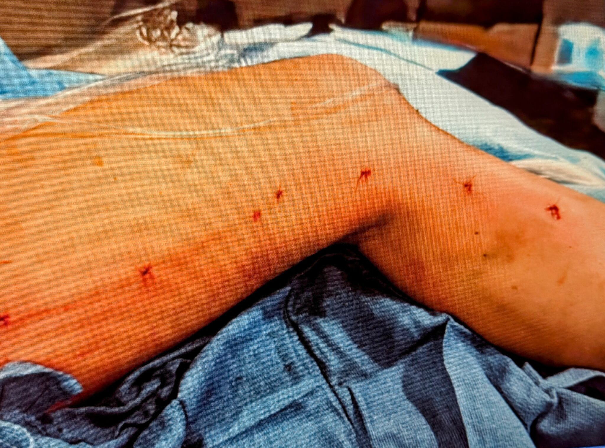
Figure 2. Immediate post-excision appearance showing multiple small incisions
required to remove a densely scarred, very superficial thrombosed segment.
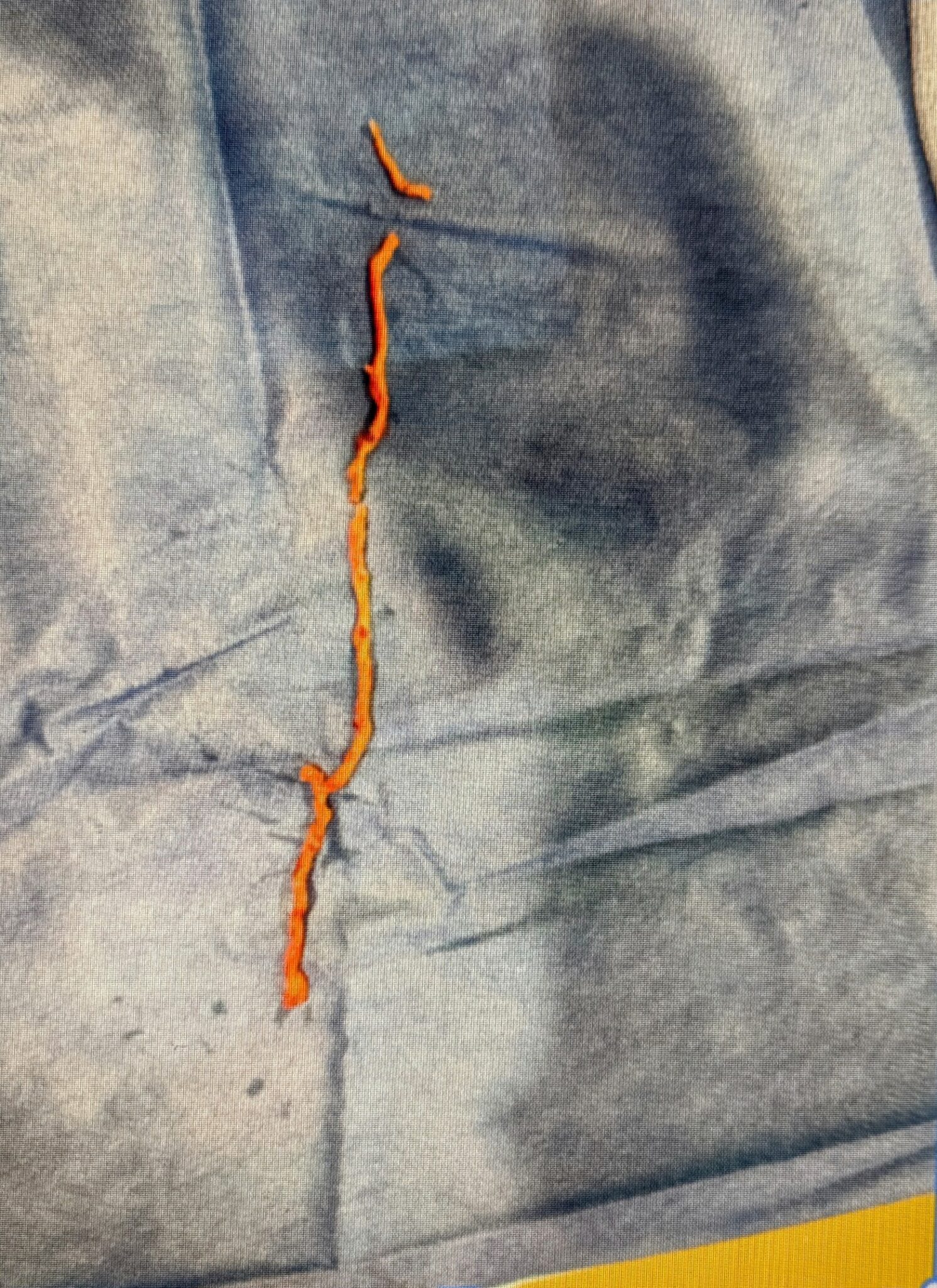
Figure 3. Back-table specimen: removed thrombosed superficial vein segment
following revision microphlebectomy/excision.
Request an Appointment
Please take a moment and fill out your request below and one of our staff members will be in contact within 24-48 business hours. If this is an emergency, call 911 immediately. If this is a non-emergent concern, please call the office Monday-Friday between 8am – 5pm at: 239-694-8346